Osteoporosis
Notes
Overview
Osteoporosis is characterised by low bone mass and micro-architectural deterioration of bone with resulting fragility and fracture risk.
Osteoporosis can be defined as a bone mineral density (BMD) of 2.5 standard deviations below the mean peak mass (for an average young healthy adult). With the exception of fracture, osteoporosis is an asymptomatic condition. The weaker bone predisposes patients to what are termed 'fragility fractures'. This refers to those that result from 'low-energy' trauma that would not normally result in fracture. Common fragility fractures include vertebral crush fractures and those of the distal wrist and proximal femur.
A number of therapeutic options are available to help prevent bone loss, including bisphosphonates and denosumab. However, the prevention of fragility fractures requires a far more holistic approach that involves optimisation of modifiable risk factors, medication review and fall prevention.
Epidemiology
Post-menopausal women are at the greatest risk of osteoporosis.
The prevalence of osteoporosis increases with age and is higher in women following menopause as oestrogen levels fall. White people are also at increased risk when compared to other ethnicities.
An ageing population is contributing to a rise in fragility fractures and it is estimated that over 500,000 occurred in 2010 in the UK (Svedbom et al).
Pathogenesis
Osteoporosis occurs due to an imbalance of bone breakdown and bone formation.
Three of the most important cell types found in bone are:
- Osteoblasts: are responsible for bone formation.
- Osteocytes: are mature cells (derived from osteoblasts) that help to maintain bone.
- Osteoclasts: are primarily responsible for bone breakdown.
As we age, the activity of osteoclasts increases and is not matched by osteoblasts. As such bone mass decreases. The ‘peak mass’ we reach as young adults is key, and a higher peak is somewhat protective. Genetics have a significant influence over the peak reached.
Oestrogen is key to the activity of bone cells with receptors are found on osteoblasts, osteocytes, and osteoclasts. Following menopause, its deficiency leads to an increased rate of age-related bone loss. This affects both the cancellous (spongy) and cortical (compact) bone.
Glucocorticoids are a commonly used medication that can lead to osteoporosis by causing increased turnover of bone. Prolonged use can result in a reduced turnover state (less bone breakdown) though even here synthesis (bone formation) is affected more leading to a loss of bone mass.
Fracture risk
Risk factors for fractures can be BMD dependent or BMD independent.
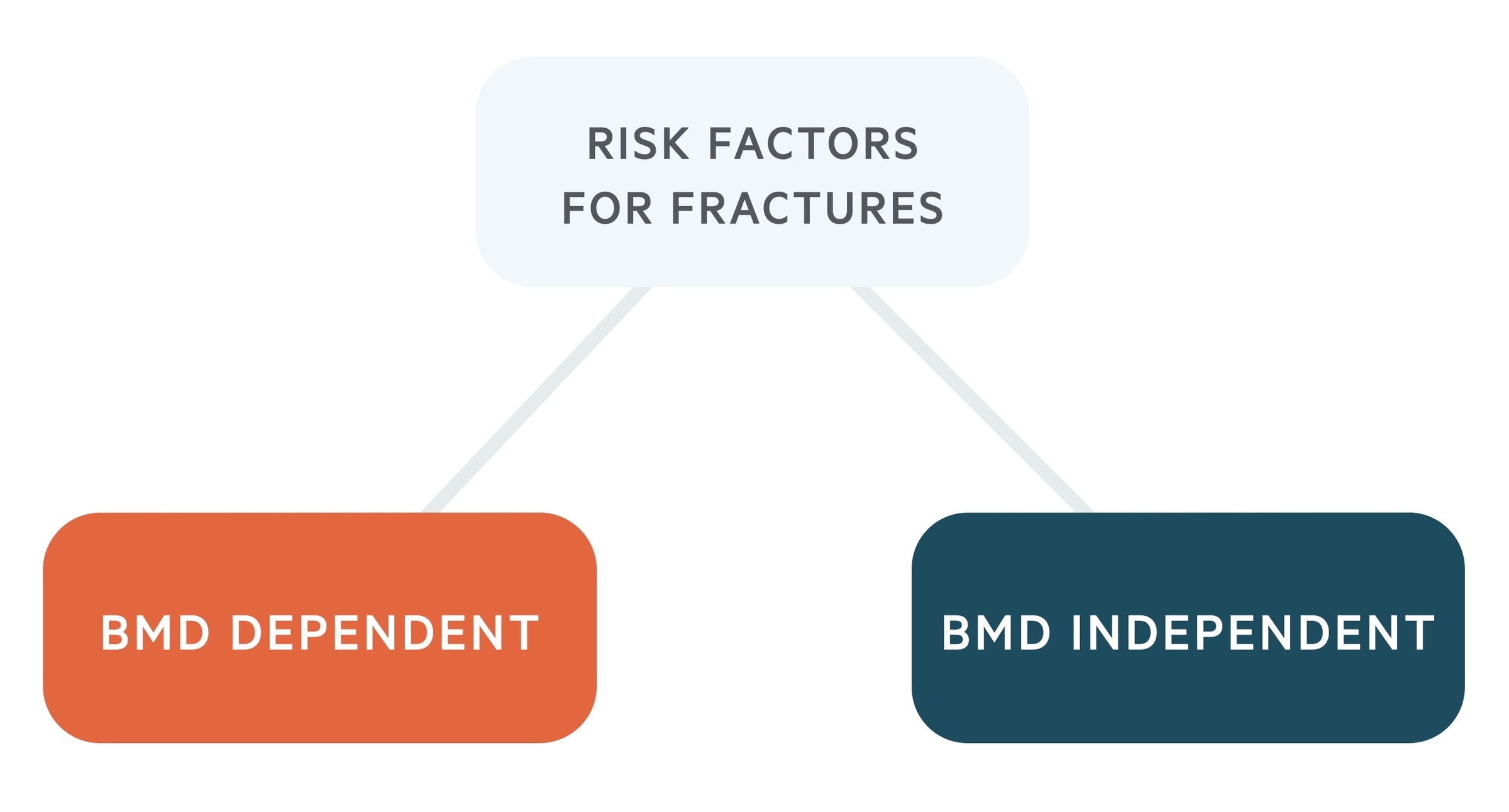
Risk factors that are BMD dependent result in an increased risk of fracture based purely on their effect on BMD. BMD independent factors increase the risk of fracture, even in the absence of reduced BMD.
BMD dependent
- Female sex
- Caucasian/Asian
- Chronic kidney disease
- Chronic liver disease
- Vitamin D deficiency
- Endocrine disorders (diabetes, Cushing syndrome, hyperparathyroidism, hyperthyroidism)
- Multiple myeloma
- Osteogenesis imperfecta
BMD independent
- Age
- Previous fragility fracture
- Family history of hip fracture
- Corticosteroids
- Alcohol (3 units or greater / day)
- Smoking
- Rheumatoid arthritis
- Low body mass index
Who to assess
Low BMD is associated with a significantly increased risk of fracture.
It is estimated there is a two times increased risk of fracture with each 1 SD decrease in BMD. However, though BMD has a high specificity for fracture risk, the sensitivity is poor - that means many patients will have fragility fractures whilst not meeting the threshold of osteoporosis. As such additional factors must be considered.
NICE (CG146) advise the following patient groups are considered for fracture risk assessment:
- All women aged 65 years and over and all men aged 75 years and over
- Women aged under 65 years and men aged under 75 years in the presence of risk factors. Examples include:
- Previous fragility fracture
- History of falls
- Family history of hip fracture
- Current use or frequent recent use of oral or systemic glucocorticoids
- Low BMI (< 18.5)
- Other causes of secondary osteoporosis
- Smoking
- Alcohol intake of more than 14 units per week for men and women.
Those aged 50 years or younger can be screened if they have one of the following risk factors:
- Current or frequent use of oral corticosteroids.
- Untreated premature menopause.
- A previous fragility fracture.
Those aged 40 years or younger may be screened if they have one of the following risk factors:
- Current or recent use of high-dose oral corticosteroids equivalent to, or more than, 7.5 mg prednisolone daily for 3 months or more.
- Previous fragility fracture of the spine, hip, forearm, or proximal humerus.
- History of multiple fragility fractures.
Additionally, GPs may consider assessing patients on SSRIs, GNRH agonists, aromatase inhibitors, antiepileptics, PPIs and thiazolidinediones, particularly if other risk factors are present.
Risk assessment tools
Using FRAX or QFracture, patients fracture risk can be categorised as high, intermediate or low risk.
Use of the FRAX and QFracture tools are advised to help determine who to investigate further with a DXA. Both take into account numerous risk factors including age, gender, and certain medications (with FRAX also giving the option of adding a DXA result). Both can be used to calculate the risk of fracture over 10 years. More details about each can be found here:
The use of these tools is not mandatory. Clinicians can proceed straight to DXA in patients over the age of 50 who have a history of fragility fracture. This may also be arranged for those younger than 40 with a major risk factor.
High risk of fragility fracture
If a patient's fracture risk is above the given threshold, offer a dual-energy x-ray absorptiometry (DXA) scan:
- T-score is ≤ -2.5: pharmacological therapy is advised with bone-sparing agents. Optimise risk factors and treat any underlying conditions.
- T-score is > -2.5: optimise risk factors and treat any underlying conditions. The DXA should be repeated at an appropriate interval, normally within 2 years.
Intermediate risk of fragility fracture
In patients whose fracture risk is close to the treatment threshold and who have risk factors - that may be underestimated on the assessment tool- arrange a DXA scan. Pharmacological therapy is advised with bone-sparing agents to those with a T-score is ≤ -2.5. Optimise risk factors and treat any underlying conditions.
Low risk of fragility fracture
For people whose fracture risk is below the recommended threshold, drug treatment is not offered. Optimise risk factors and treat any underlying conditions and follow up within 5 years.
Investigations
Dual-energy X-ray absorptiometry (DXA) scans are used to assess BMD.
DXA scan
Clinicians may proceed straight to DXA in patients over the age of 50 who have a history of fragility fracture. This may also be arranged for those younger than 40 with a major risk factor.
A DXA scan utilises X-rays to assess the density of bones, commonly the hip and spine. Patients are scored using the T-score which compares their bone density to that of a young adult. The T-score represents the number of standard deviations a patient's bone density is from the average for a healthy young adult of the same gender. A negative score tells you their bone density is below this average. Based on their T-score a patient can be normal, osteopenic (a less severe reduction in bone mineral density) or osteoporotic:
- Normal: a T-score > -1
- Osteopenia: a T-score of -1 to -2.5
- Osteoporosis: a T-score of ≤ -2.5
Vertebral fracture assessment
In addition to the above assessments consider lateral lumbar and thoracic spine radiographs in patients with:
- History of ≥ 4cm height loss
- Kyphosis
- Recent or current long-term oral corticosteroids
- BMD T-score ≤ -2.5
Pathological fractures and secondary osteoporosis
In any patient with a history of fragility fractures, it is necessary to consider the possibility of a pathological fracture. The term pathological fracture is normally used to refer to fractures that occur secondary to malignancy (primary bone or metastasis).
It is also important to consider, investigate and treat secondary causes of osteoporosis where appropriate. Secondary causes include endocrine conditions (diabetes, Cushing syndrome, hyperparathyroidism, hyperthyroidism), malabsorptive conditions (e.g. IBD), chronic liver disease, COPD, CKD and certain rheumatological conditions.
Treatment
Oral bisphosphonates are normally first-line treatment for patients with osteoporosis.
Bisphosphonates
When medical therapy is indicated bisphosphonates (e.g. alendronate) are normally prescribed first-line. They may also be given to patients on corticosteroids (taking the equivalent of prednisolone 7.5 mg daily for 3 months or longer) and for the prevention of postmenopausal osteoporosis.
- Alendronate: commonly used and is licensed in postmenopausal osteoporosis and osteoporosis in men. It must be taken in the morning with a glass of water, after fasting overnight and at least 30 minutes prior any food or drink. After taking it the patient must stay upright for 30 minutes.
- Zoledronic acid: may be used in those not tolerating oral preparations. Given as an IV injection once a year in postmenopausal osteoporosis and osteoporosis in men.
Oral bisphosphonates have a number of side effects that are relatively common and include GI disturbance, oesophagitis and headaches. Contraindications include renal impairment (see BNF for individual cut-offs), hypocalcaemia, pregnancy/breastfeeding (they are only licenced for postmenopausal women) and hypersensitivity.
Osteonecrosis of the jaw may rarely occur. A dental exam is advised prior to treatment in those with dental disease, tobacco use or glucocorticoid use. Additionally, where possible dental treatment should be avoided. Good oral hygiene should be advised with regular check-ups.
Another rare complication is an atypical femoral fracture. The patient should be aware to present if they develop pain in their hip or thigh.
Long term treatment is an area of ongoing research. The risk of atypical fractures and osteonecrosis of the jaw increase with time. Review is recommended at 3-5 years, see NOGG for more information.
Denosumab
Denosumab is a monoclonal antibody against Receptor Activator of Nuclear factor Kappa B ligand (RANK ligand) given via subcutaneous injection every 6 months.
It is used in postmenopausal osteoporosis and osteoporosis in men. It can also be used for patients on corticosteroids (taking the equivalent of prednisolone 7.5 mg daily for 3 months or longer) and in the treatment of bone loss associated with hormone ablation in prostate cancer.
Side effects include cellulitis and hypocalcaemia. Monitoring of calcium may be required. It is contraindicated in hypocalcaemia and hypersensitivity and avoided in pregnancy. Rarely osteonecrosis of the jaw and atypical femoral fractures may be seen. Precautions are similar to those discussed for bisphosphonates.
Raloxifene
Raloxifene is a selective oestrogen receptor modulator and may be used in postmenopausal osteoporosis. Side effects include hot flushes, vaginal dryness and leg cramps.
There is a risk of venous thromboembolism and stroke. It should not be used in women with a history of venous thromboembolism or if a patient has prolonged immobilisation. Contraindications also include cholestasis, endometrial cancer, undiagnosed uterine bleeding, child-bearing age and lactation. It has been associated with a reduced risk of breast cancer but use in patients with breast cancer should be avoided.
Hormone replacement therapy (HRT)
HRT can be given in the form of unopposed oestrogen or oestrogen in combination with progesterone for the prevention of fracture in women at high risk. It is normally reserved for use in younger women as the side effect profile is better.
Side effects are numerous (and dependent on the preparation) and include breast tenderness, leg cramps and changes to mood. There also appears to be an increased risk of blood clots, stroke, endometrial cancer (oestrogen only preparations) and breast cancer.
Lifestyle factors
Discuss adverse lifestyle factors like alcohol and smoking. Provide the necessary support to help patients who are considering a lifestyle change. Assess vitamin D and calcium and consider the need for supplementation.
A falls assessment should be completed (including medication review) - and steps are taken to reduce the risk of falls. Regular ‘weight bearing’ exercise, taking into account individual patients abilities, should be advised.
Last updated: October 2024
- NOGG 2017: Clinical guideline for the prevention and treatment of osteoporosis
- QFracture
- FRAX
Further reading
.
Have comments about these notes? Leave us feedback