Perthes’ disease
Notes
Introduction
Perthes' disease affects the hip joint in children and is characterised by idiopathic avascular necrosis of the femoral epiphysis and resulting structural changes.
Fully titled Legg–Calvé–Perthes disease after three physicians who independently and simultaneously described the condition, it most commonly presents in children aged 3-12 with features of atraumatic groin/hip pain, limp and stiffness. Though normally unilateral, up to 10-20% of cases are bilateral. The aetiology of Perthes’ disease is not yet understood.
As described above it is characterised by idiopathic avascular necrosis of the femoral epiphysis. It is important to have a firm understanding of these terms:
- Idiopathic: means the underlying cause of the condition is unknown.
- Avascular necrosis: refers to the death of bone tissue secondary to an impaired blood supply.
- Femoral epiphysis: refers to the (proximal) end of the femur, separated from the metaphysis by the growth plate.
It is a rare condition, estimated to affect around 1 in 9000 children. Management is based around symptomatic relief and physiotherapy with the goal of maintaining normal hip anatomy and function. Though most children will not require it, a proportion will need surgical intervention.
Epidemiology
Perthes’ disease is estimated to affect 1 in 9000 children.
The condition is more common in males compared to females (approx. 4:1) and is seen primarily in Caucasians.
It tends to occur between the ages of 3 to 12 with incidence peaking between 4 and 8.
Risk factors
Perthes’ disease is more commonly seen in males and those of North European descent.
There are a number of risk factors associated with the development of Perthes’ disease, these include:
- Male gender
- Caucasian ethnicity
- Passive smoking
- Deprivation
- Obesity
- Prenatal factors (e.g. low birth weight)
Pathology
Perthes’ disease is characterised by idiopathic avascular necrosis of the hip in children.
Avascular necrosis refers to the disrupted blood supply of bone tissue with resulting necrosis. The cause of this remains unknown. There are a number of theories including the repetitive microtrauma hypothesis.
The course of the disease is variable but with time revascularisation occurs and necrotic bone is replaced. In the absence of appropriate management, there is a high risk of persisting femoral head deformity, leg length discrepancy and reduced function.
Clinical features
Children most commonly present with atraumatic hip pain and a limp.
Symptoms normally develop with a gradual and insidious onset and are bilateral in 10-20% of cases. The pain may be in the hip or groin and can be felt as referred pain to the knee. Activity tends to worsen the pain but it is often present at rest as well.
A limp caused by pain (antalgic gait) is common. In later disease a Trendelenburg gait - reflecting weakness of the hip abductors - can be seen.
Early in the disease process examination may be relatively normal but as it progresses it often reveals a reduction in the range of movement, particularly abduction and internal rotation of the affected hip.
Differential diagnosis
There are a number of alternative diagnoses that should be considered.
- Septic arthritis
- Slipped Capital Femoral Epiphysis (SCFE)
- Transient synovitis
- Other causes of avascular necrosis:
- Renal failure
- Sickle cell disease
- Gaucher disease
- Glucocorticoid use
- Lupus
- HIV
Imaging
Pelvic X-rays and MRI hips are used to diagnose and monitor Perthes’ disease.
X-ray
Pelvic/hip X-rays (anterior-posterior or AP and frog-leg lateral views) are the first-line imaging investigations. Early on in the disease process changes may be subtle or not seen.
As the disease develops reduction in size and lucency of the epiphysis is seen. Evidence of a joint effusion may be present. Later fragmentation of the epiphysis can occur.
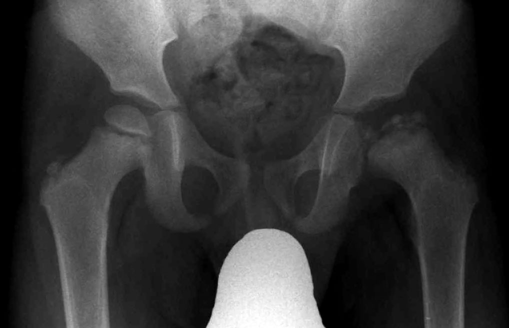
Fragmentation and destruction of left hip epiphysis
MRI
An MRI has the advantage of showing early changes, prior to them appearing on an X-ray. It also avoids radiation. It is however expensive, time-consuming and can be distressing for children.
Management
Symptomatic relief and physiotherapy form the mainstay of management.
There is a lack of widespread consensus on how to best manage Perthes’ disease. This reflects our incomplete understanding and conflicting research. The general aim is to reduce pain and avoid irreversible damage and deformity to the femoral head.
Techniques may include restricted weight-bearing activities and range of motion exercises under the guidance of a specialised physiotherapist. The psychological impact of any restriction in normal activity should not be discounted and support offered where appropriate.
Patients require monitoring to assess for disease progression or signs of healing and resolution. A proportion can need surgical intervention, particularly older patients with later-stage disease. Techniques include surgical osteotomy, where the alignment of the joint is improved to allow better remodelling of the femoral head.
Prognosis
Most children recover well with conservative measures, though there is a risk of early osteoarthritis later in life.
Age is a significant prognostic factor. Children affected at an older age (e.g. upwards of 8) tend to have a poorer outcome. Other negative prognostic factors include female gender, obesity and stiffness on examination.
In the long term, there is a higher incidence of early hip osteoarthritis in patients affected by Perthes’ disease, a risk associated with any residual structural abnormality.
Last updated: July 2021
Have comments about these notes? Leave us feedback