Biliary colic
Notes
Overview
Biliary colic refers to a pain in the RUQ/epigastrium caused by gallstones.
It is the most common symptomatic manifestation of cholelithiasis (gallstones) affecting around 10-20% of patients. Though termed a 'colic' the pain is normally constant lasting from 30 minutes to 6 hours.
The pain occurs when a stone impacts against the cystic duct during contraction of the gallbladder with increased pressures in the gallbladder itself. Biliary colic is generally considered an indication for elective laparoscopic cholecystectomy after appropriate investigation.
See our Cholelithiasis notes for more about the aetiology, types and risk factors.
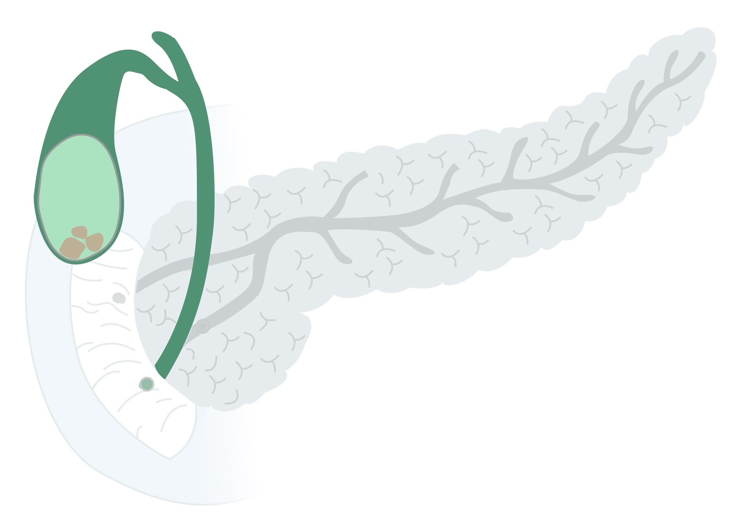
Gallstones
Cholelithiasis (gallstones) refers to the development of a solid deposit or ‘stone’ within the gallbladder.
Though largely asymptomatic in a significant proportion of patients they become problematic. In the UK around 60,000 cholecystectomies are performed each year.
Gallstones affect up to 20% of the population. The prevalence of gallstones increase with advancing age before levelling off in the sixth - seventh decade of life. They are more common in women and tend to affect those of caucasian, Native American and hispanic backgrounds more.
Outside of haemolytic anaemias, gallstones are rarely seen in childhood. The vast majority of people with gallstones will remain asymptomatic (80%). There are a number of risk factors are associated with the development of cholelithiasis:
- Age
- Female sex
- Genetic predisposition
- Obesity
- Rapid weight loss / prolonged fasting
- Diabetes
- Medications (e.g. oestrogen replacement therapy, ceftriaxone, octreotide)
- Crohn's disease
- Diet (high in triglycerides, refined carbohydrates)
Increased red cell turnover is implicated in the development of black pigment stones specifically.
Clinical features
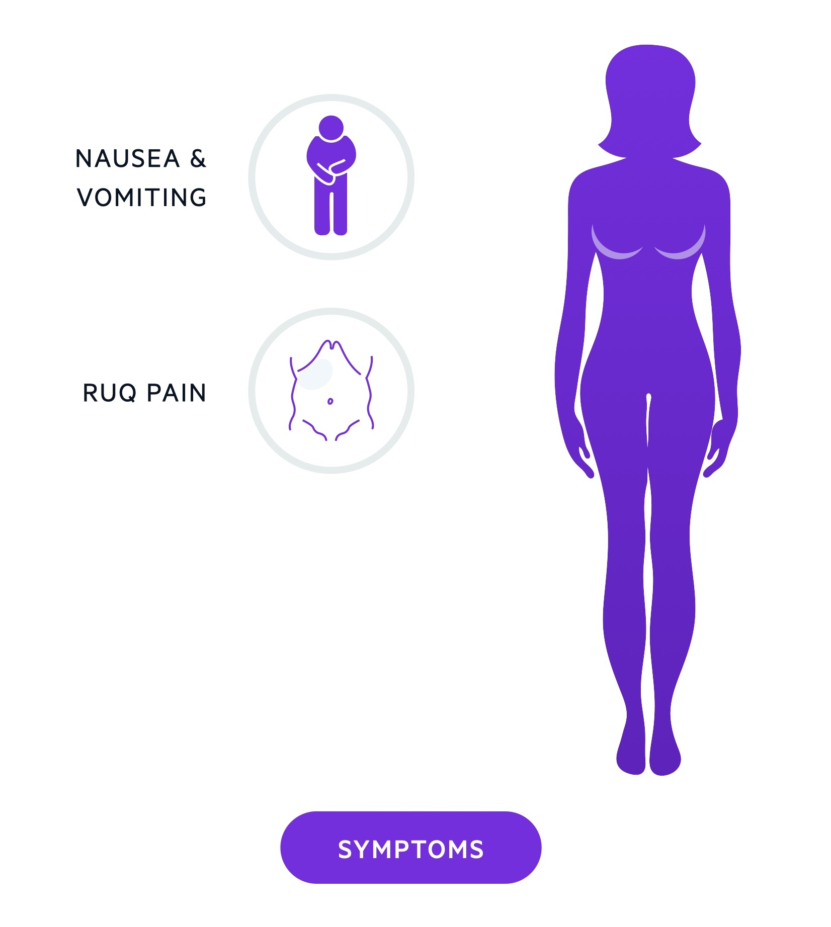
Biliary colic is a self-limiting pain in the epigastrium/RUQ.
Though termed 'colic' the pain is normally constant with episodes typically lasting 30 minutes - 6 hours. It is frequently associated with nausea and may precipitate vomiting.
- Intermittent RUQ/epigastric pain
- Nausea / vomiting
Investigations
An USS abdomen is highly sensitive for the diagnosis of gallstones.
Patients with suspected gallstones should have an abdominal USS and LFTs arranged:
- USS: An abdominal USS is an excellent non-invasive, radiation free investigation for gallstones. Of note, though an USS may identify gallstones - it cannot guarantee the pain experienced by the patient has been caused by the stones. It can also assess the CBD for dilation and sometimes identify stones in the CBD.
- LFTs: Liver function tests represent an essential component of the work-up. Derangement may be indicative of stones within the biliary system (which can be asymptomatic). It is essential to identify patients with CBD stones prior to any cholecystectomy. See the management section for more details.
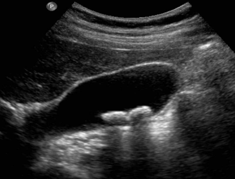
Gallstones seen within the gallbladder on USS
Image courtesy of Dr Andrew Dixon and Radiopaedia
Many other investigations may be ordered depending on the presentation and co-morbidities. Alternative diagnoses should be considered and appropriately investigated. Gastritis and peptic ulcer disease may present with similar symptoms - consider an upper GI endoscopy if diagnostic uncertainty exists.
Management
Biliary colic is managed with symptomatic relief and elective cholecystectomy.
Symptomatic management and prevention
Conservative measures may be used to treat acute symptoms and reduce episodes of colic:
- Analgesia: Simple pain relief with paracetamol and NSAIDs (in the absence of contra-indications). Occasionally opioid analgesia may be required.
- Diet: A low-fat diet may be trialled with some patients experiencing a significant reduction in the number and severity of episodes.
Surgical management
Typically once gallstones have become symptomatic, they are likely to be troublesome in the future. For most patients an elective cholecystectomy is indicated.
A routine general surgery review with a view to considering operative management should be arranged. In patients with significant co-morbidities, a cholecystectomy may represent unacceptable risk and as such conservative measures trialled.
Prior to cholecystectomy it is key that CBD stones are excluded. All patients will have had an USS and set of LFTs as a minimum. If there is suspicion of CBD stones (e.g. dilated CBD or CBD stone on USS, raised bilirubin on LFTs) then there are two main options:
- MRCP +/- ERCP: MRCP allows for confirmation of stones in the biliary tree. If present ERCP allows for therapeutic intervention with stone retrieval, sphincterotomy and stent placement prior to cholecystectomy.
- On-table cholangiogram: Less commonly available and technically challenging. During the laparoscopic cholecystectomy the bile duct is intubated to allow the injection of dye with fluoroscopy in-theatre to diagnose stones in the biliary tree. Various techniques may then be used to retrieve/expel stones.
Last updated: May 2021
Have comments about these notes? Leave us feedback