Elbow fractures
Notes
Definition
An elbow fracture is a bony injury to one or more of the three bones comprising the elbow joint: the distal humerus, proximal ulna and proximal radius.
Elbow fractures are common and have a bimodal distribution, generally sustained from a fall directly onto the elbow or outstretched hand. They are frequently associated with ligamentous injury and sometimes with elbow dislocations.
Any patient with an elbow fracture must be carefully assessed for injury to the nerves and blood vessels around the joint.
Broadly, there are three main types of elbow fractures:
- Distal humerus and supracondylar fractures
- Olecranon fractures (part of the proximal ulna)
- Radial head and neck fractures
Anatomy
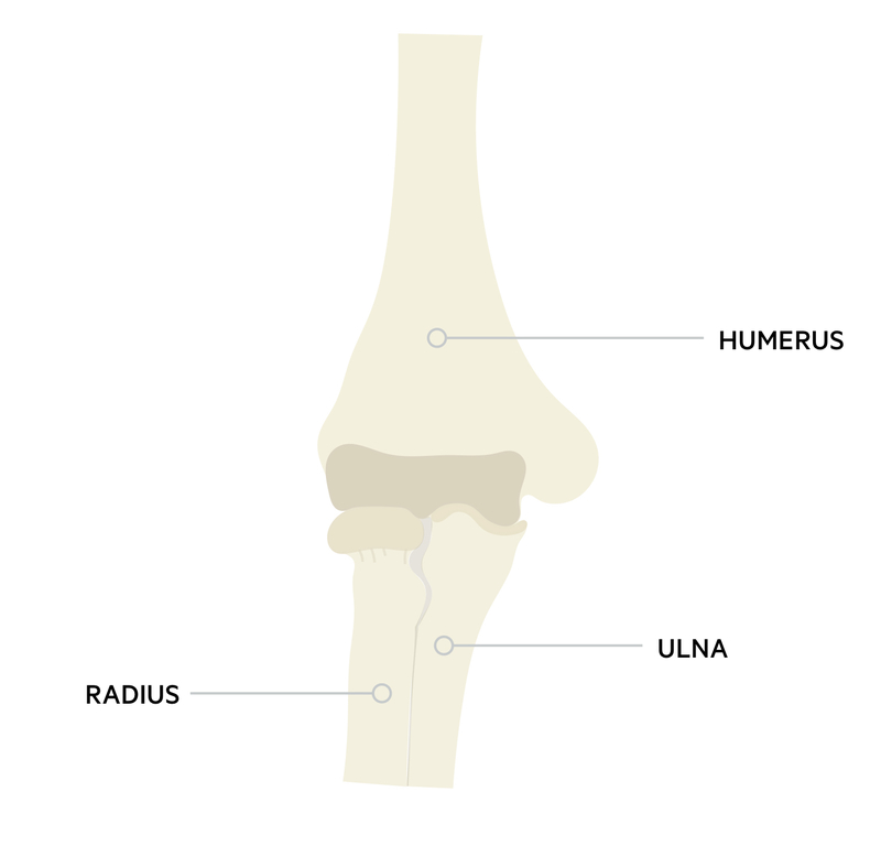
The elbow is a compound synovial joint comprised of three articulations between the humerus, radius and ulna.
The elbow permits flexion and extension, in addition to pronation and supination of the forearm. There are three articulations:
- Humeroulnar joint - a hinge joint between the trochlea of the humerus and the ulna.
- Humeroradial joint - between the capitellum of the humerus and the radial head.
- Proximal radioulnar joint - a pivot joint between the radial head and proximal ulna.
NB - The proximal radioulnar joint is sometimes considered separately to the elbow joint, but is within the same joint capsule.
Ligaments
The elbow joint is stabilised by strong ligaments on each side. The ulna collateral ligament (UCL) runs medially and the radial collateral ligament (RCL) laterally. There is also an annular ligament which runs around the radial head to support the radioulnar joint.
Blood vessels
The brachial artery descends anteriorly from the arm and divides into the radial and ulnar arteries usually around the level of the radial neck. From these arteries, there is a rich network of blood vessels anastomosing around the elbow. The brachial artery is susceptible to injury in supracondylar fractures of the humerus.
Nerves
The median, radial and ulnar nerves descend from the brachial plexus and cross the elbow joint.
- Median nerve - runs medially to the brachial artery in the cubital fossa in the anterior elbow.
- It gives off an important motor branch called the anterior interosseous nerve before continuing into the forearm.
- Radial nerve - runs anteriorly to the lateral epicondyle of the humerus.
- It then divides into superficial and deep (posterior interosseous) branches.
- Ulnar nerve - passes posteriorly to the medial epicondyle of the humerus.
Distal humerus fractures
Distal humerus fractures in adults and supracondylar humerus fractures in children should be considered separately.
This injury is uncommon in adults. They are most commonly seen in older patients with osteoporotic bone or following high energy injuries in younger adults.
Fractures are often very unstable and there is a high risk of neurovascular injury. Surgery is often required and subsequent elbow stiffness very common.
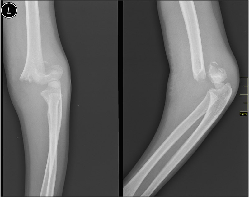
A distal humerus fracture. Image courtesy of Nevit Dilmen
Supracondylar humerus fractures
Supracondylar fractures are common in children and result from falling on an outstretched hand.
Displaced fractures are associated with injury or kinking of the brachial artery and median nerve (anterior interosseous branch).
A child with a supracondylar fracture and an ischaemic arm requires immediate reduction and operative fixation of the fracture. If this does not result in reperfusion of the limb, the brachial artery must be explored.
The Gartland classification is commonly used to describe supracondylar fractures in terms of degree of displacement:
- Type 1: Undisplaced or minimally displaced
- Type 2: Displaced, with intact cortex
- Type 3: Completely displaced
A supracondylar fracture. Image courtesy of Nevit Dilmen
Have comments about these notes? Leave us feedback
